| Female Pelvis, Page 2 | ||||
|
||||
|
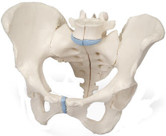
3-Part Female Pelvis This life size three part model represents an original cast of a bony female pelvis and shows all the details of the following anatomical structures: Two hip bones, the pubic symphysis, the sacrum and the coccyx, the fifth lumbar vertebra with intervertebral disc. A midsagital section through the fifth lumbar vertebra, sacrum and coccyx, allow both halves of the pelvis to be disassembled revealing a part of the cauda equina in the vertebral canal. The left half of the fifth lumbar vertebral body is removable. |
||||
|
||||
|
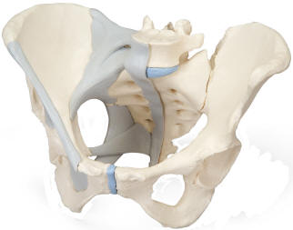
3-Part Female Pelvis with Ligaments Same as above, but the right half of the model also shows the following pelvic ligaments: inguinal ligament, sacrotuberous ligament, sacrospinous ligament, anterior sacroiliac ligaments, iliolumbar ligament, anterior longitudinal ligament, interosseous sacroiliac ligament, posterior sacroiliac ligament and obturator membrane. |
||||
|
||||
|
|
||||
|
||||
|
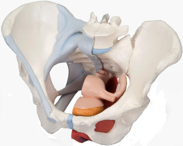
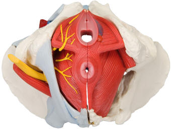
4-Part Female Pelvis This life size four part model of a female pelvis represents detailed information about the topography of bones, ligaments, pelvic floor muscles and female pelvic organs. The right half shows the bones with pelvic ligaments. In addition, the left half of the pelvis contains the muscles of the pelvic floor including levator ani, ischiocavernosus, deep and superficial transverse perineal, external anal sphincter, external urethral sphincter. A partially removable bulbospongiosus demonstrates the vestibular bulb and Bartholin gland. The removable midsagital section through the urinary bladder, vagina, uterus and rectum demonstrates the relationship to the muscles of the pelvic floor within its openings for urethra, vagina and rectum. The female genital organs are detailed for gynecological and other anatomy studies. |
||||
|
||||
|
|
||||
|
||||
|
|
||||
|
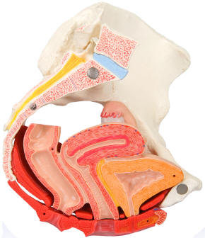
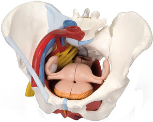
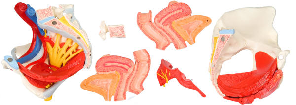
6 Part Female Pelvis This life size six part model of a female pelvis represents detailed information about the topography of bones, ligaments, vessels, nerves, pelvic floor muscles and female genital organs. It presents the whole pelvic floor with partially removable midsagitally sectioned external anal sphincter, external urethral sphincter, deep and superficial transverse perineal and bulbospongiosus. The rectum, uterus with fallopian tubes, ovaries and vagina are also removable and can be disassembled into both halves by midsagital section. The right pelvic half demonstrates the divisions and topographical anatomy of the common iliac artery, the external and internal artery and also of the common iliac vein and the external iliac vein. The right sacral plexus, right sciatic nerve and right pudendal nerve are also shown. Bones and ligaments presented: Two hip bones, the pubic symphysis, the sacrum and the coccyx, the fifth lumbar vertebra with intervertebral disc. A midsagital section through the fifth lumbar vertebra, sacrum and coccyx, allow both halves of the pelvis to be disassembled revealing a part of the cauda equina in the vertebral canal. The left half of the fifth lumbar vertebral body is removable. The right half of the model shows the following pelvic ligaments: inguinal ligament, sacrotuberous ligament, sacrospinous ligament, anterior sacroiliac ligaments, iliolumbar ligament, anterior longitudinal ligament, interosseous sacroiliac ligament, posterior sacroiliac ligament and obturator. |
||||
|
||||
|
|
||||
|
|
||||
|
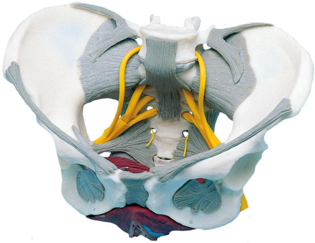
Premier Female Pelvis This highly-detailed life-size model of the female pelvis shows the hip bone, muscles, ligaments and the main nerves. The 2-part pelvic floor is removable. Learning the anatomy of the female pelvis is easy with this model. |
||||
|